RadpicsAI logo
  • Home
  • About Us
  • Viewer
  • Features
  • Plans
Get Started

Desktop + Mobile

Advanced DICOM Viewer
for Desktop & Mobile

View, analyze and work with medical images with a powerful, intuitive DICOM viewer built for modern imaging workflows.

Explore PlansSee the Viewer

View•Analyze•Measure

radpicsai.com / viewer
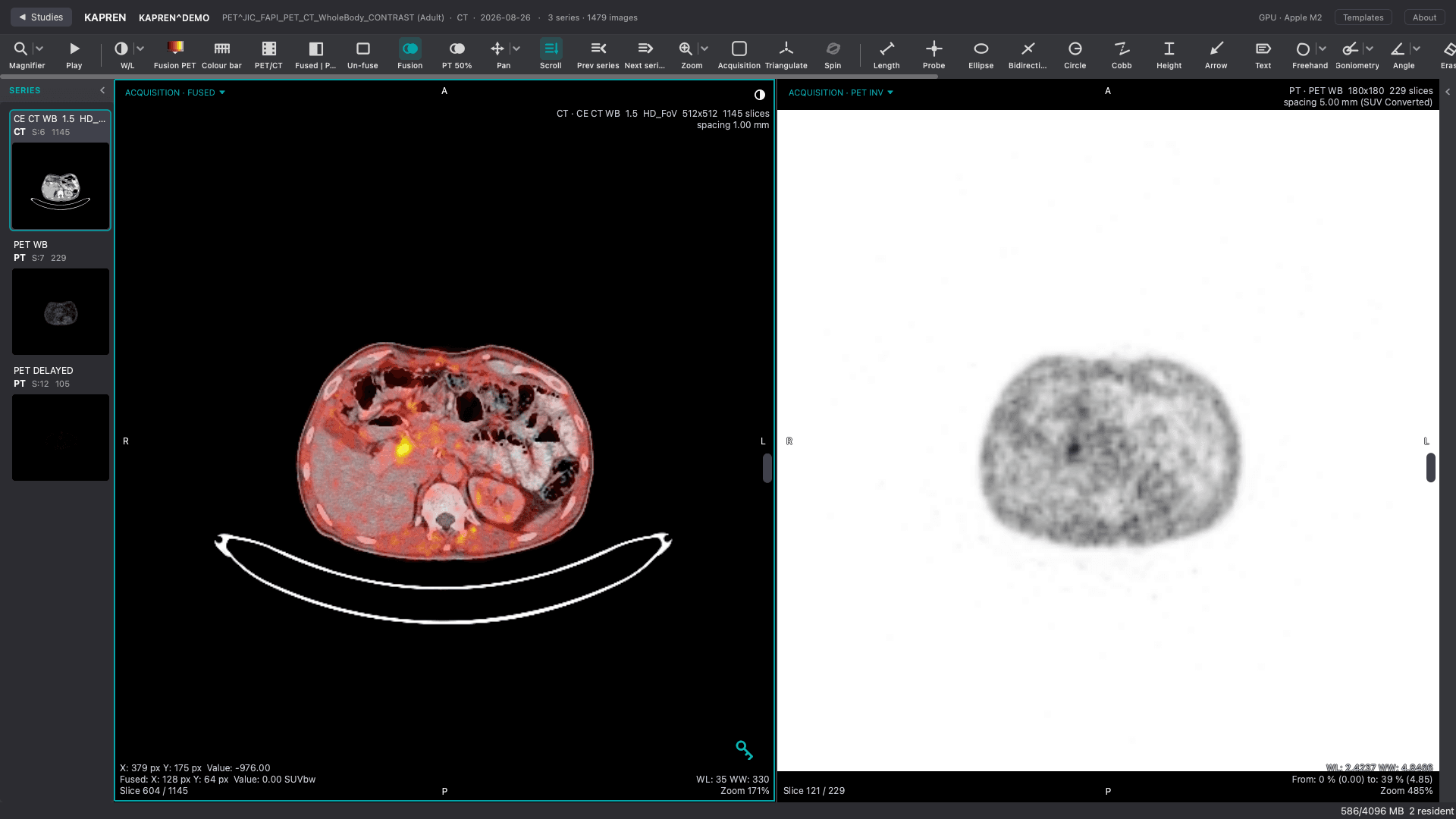
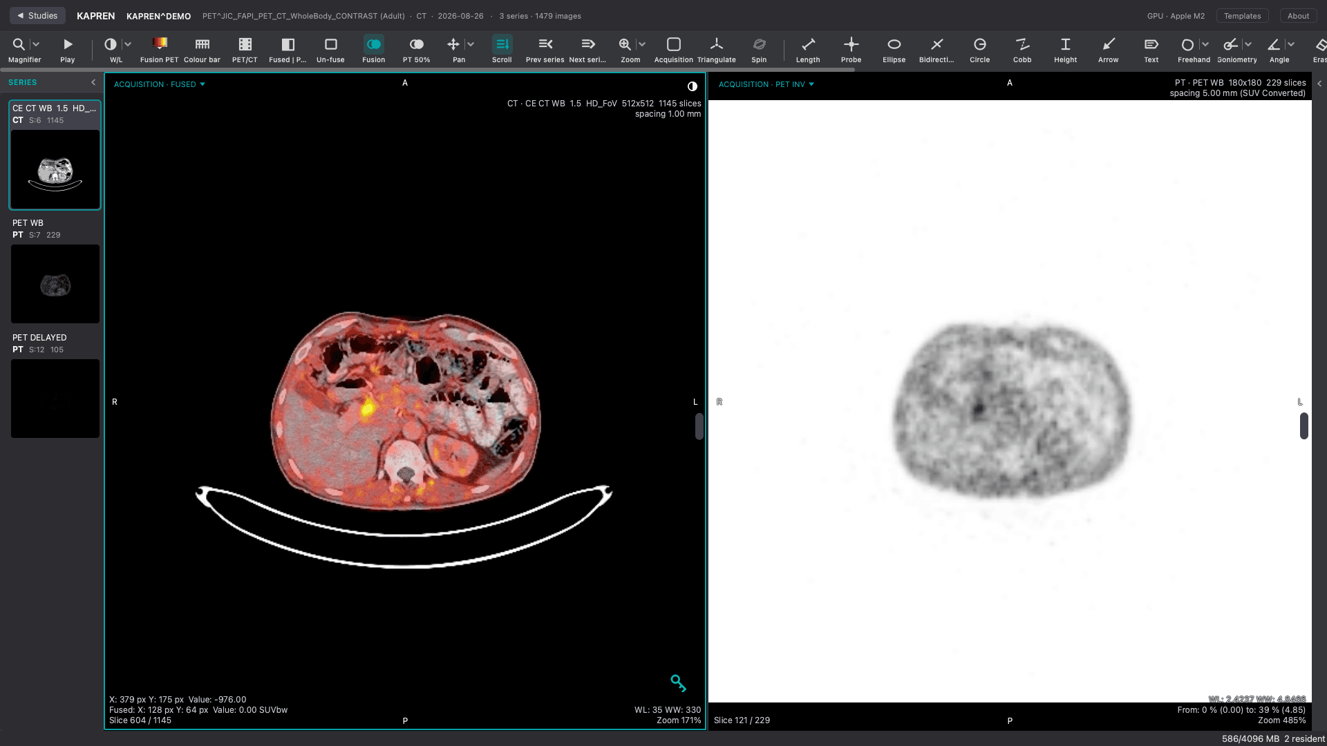
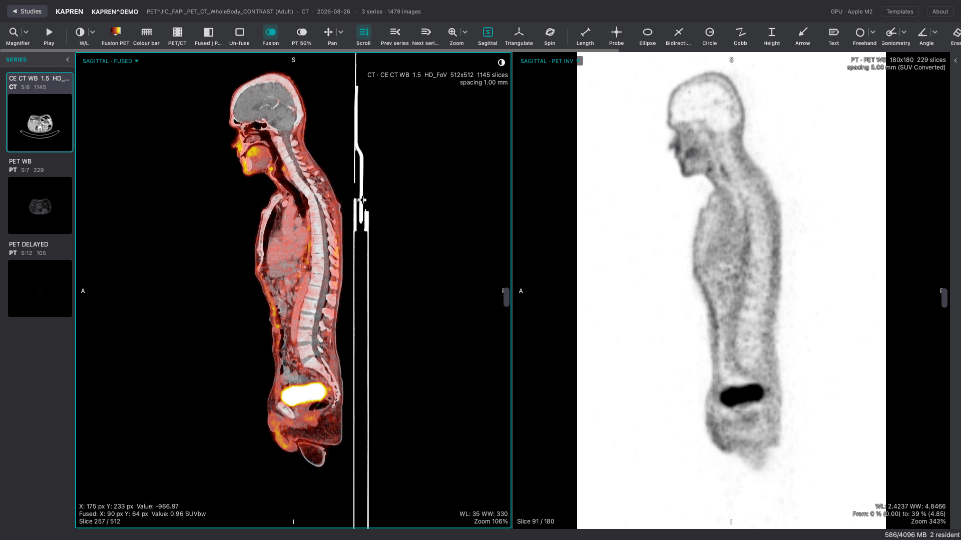
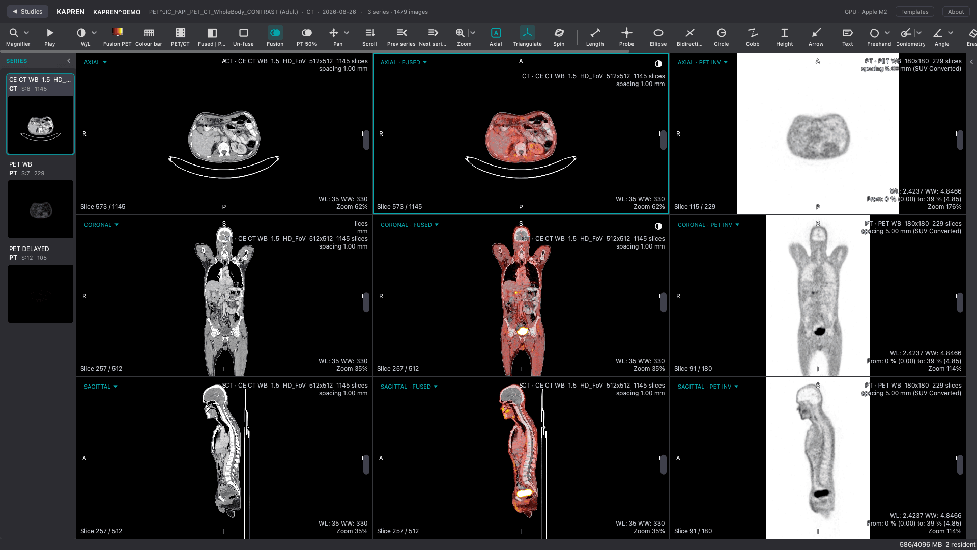
Desktop DICOM viewer — study browser, 2×2 viewport, toolbar
Mobile DICOM viewer

Viewer

One Viewer. Multiple Devices.

radpicsai.com / viewer
Desktop viewer — study browser, series thumbnails, 2×2 viewport, toolbar

Built for the reading room

A full multi-viewport workspace with the tools a radiologist reaches for on every study.

  • Multi-viewport
  • Series navigation
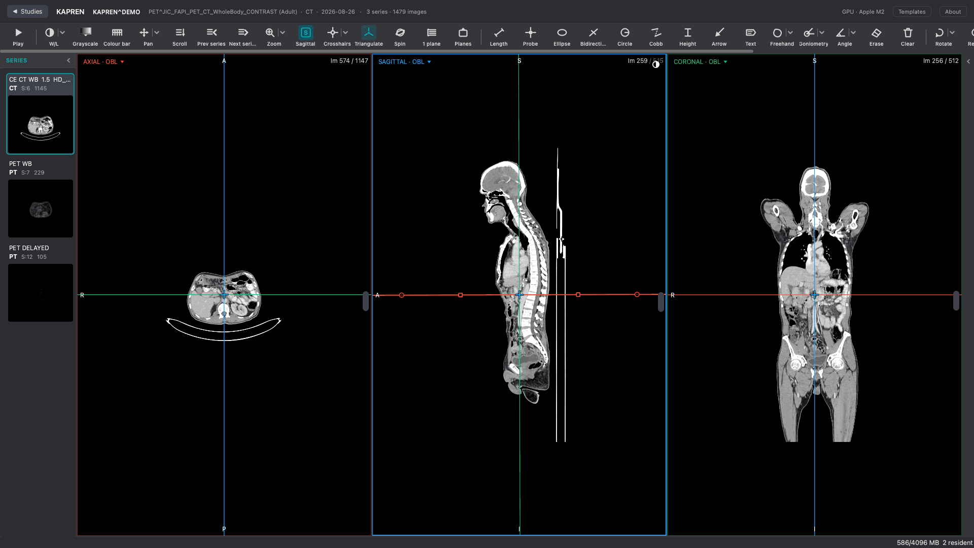
  • MPR
  • MIP
  • Measurements
  • Annotations

Features

Everything You Need in a DICOM Viewer

Core viewing, advanced visualization, measurements, annotation and comparison — grouped the way you actually work.

Core Viewing

Everything needed to open a study and read it.

  • DICOM

    Open and render standard DICOM studies.

In the viewer

See Each Tool at Work

radpicsai.com / viewer
Measurements — Length, angle, area and ROI measurements taken directly on the image.

Length, angle, area and ROI measurements taken directly on the image.

Plans

Choose the Plan That Fits Your Workflow

Every plan includes the full viewer. What changes is how many people use it — and how far the mobile app and AI go.

Base

1 user

For a small reading team working across devices.

₹1,999/month

  • Every viewer feature included
  • Desktop viewer — unlimited studies
  • Measurements & annotations
  • MPR
  • MIP and 3D
  • Mobile app included

Mobile app limited to 3 DICOM views per day

Most popular

Pro

3 users

For larger teams that want AI in the reporting loop.

₹4,999/month

  • Everything in Base
  • 3 user accounts
  • Unlimited mobile app viewing
  • Desktop viewer — unlimited studies
  • Priority support

Enterprise

More than 3 users

For larger teams that want AI in the reporting loop.

Custom

  • Everything in Pro
  • Unlimited user accounts
  • AI report generation
  • Advanced AI features
  • Custom integrations & onboarding
  • Dedicated support

Prices in INR, per month. Taxes extra.

Enquiry

Still Have Questions?

Send us your details and we’ll get back to you — no payment needed.

RadpicsAI logo

AI-powered PACS software, DICOM viewing, and radiology reporting — to connect hospitals, diagnostic centers, and remote radiologists.

Book Free Demo

Explore

  • Home
  • About Us
  • Solutions
  • Services
  • Features

Head Office

RadpicsAI Technologies Pvt. Ltd.

601, 6th Floor, Rupa Solitaire,Millennium Business Park,Plot No. A-1, Mahape,Navi Mumbai - 400710

Contact

  • +91 88288 41603
  • contact@radpicsai.com

Download App

QR code to download the RadpicsAI mobile app

Scan to download the RadpicsAI mobile app.

Connect With Us
  • Privacy Policy
  • Contact Us

©2025-26 RadpicsAI, all rights reserved

Back to top

Series Navigation

Move through series and slices at speed.

  • Zoom

    Magnify any region without losing detail.

  • Pan

    Reposition the image inside the viewport.

  • Window / Level

    Tune contrast and brightness to the tissue.

  • Rotate

    Rotate the image to the preferred orientation.

  • Flip

    Mirror horizontally or vertically.

  • Invert

    Invert greyscale for alternate contrast.

  • Cine

    Play multi-frame series as a loop.

  • Advanced Visualization

    Move past a single plane.

    • MPR

      Multiplanar reconstruction for detailed anatomical visualization.

    • MIP

      Maximum intensity projection for high-density structures.

    • 3D

      Interactive volumetric visualization.

    • Multi-Planar Viewing

      Axial, coronal and sagittal side by side.

    • Crosshair Synchronization

      Lock anatomical position across viewports.

    Measurements

    Quantify what you are looking at.

    • Length

      Point-to-point distance measurement.

    • Angle

      Measure angles between structures.

    • Area

      Measure enclosed regions.

    • ROI

      Region-of-interest statistics.

    • Pixel Measurement

      Read individual pixel values.

    Annotation

    Mark findings and hand them on.

    • Arrow

      Point directly at a finding.

    • Text

      Add free text to the image.

    • Freehand

      Trace irregular boundaries.

    • Shapes

      Rectangles, ellipses and polygons.

    • Labels

      Name structures and findings.

    • Markers

      Drop reference markers on a slice.

    Comparison

    Read studies against each other.

    • Multi-Viewport

      Several viewports in one workspace.

    • Side-by-Side Comparison

      Place two images next to each other.

    • Series Comparison

      Compare series within a study.

    • Prior Study Comparison

      Bring up a previous study alongside.